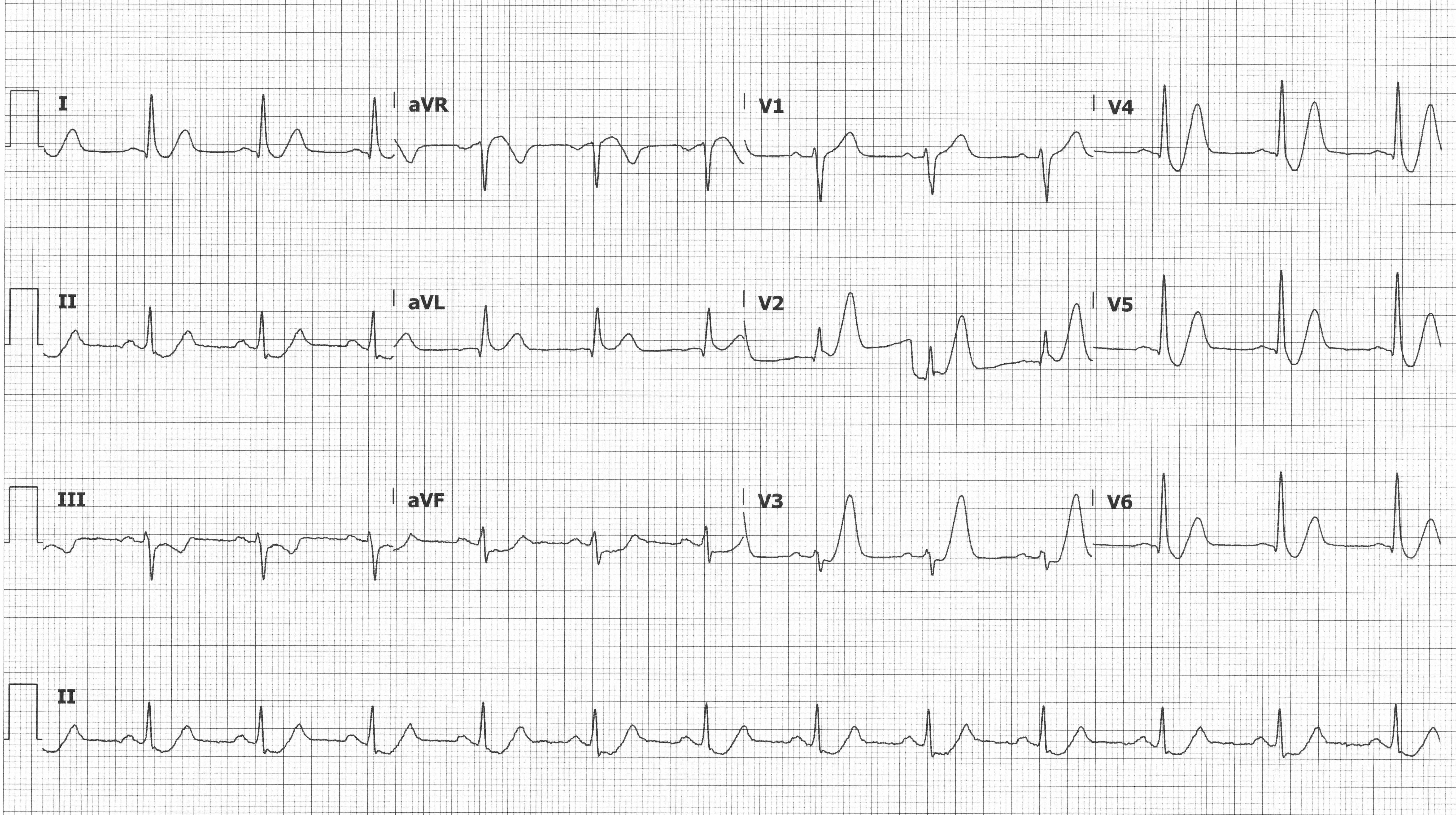
Nstemi Ekg - Myokardinfarkt: STEMI vs. NSTEMI | Online Medizin lernen / The standard 12 lead electrocardiogram (ecg) has several limitations.. Ecg which does not meet the criteria for stemi or stemi equivalent and. This video is part of the complete ekg. There is atrial fibrillation at a rate of 95. The standard 12 lead electrocardiogram (ecg) has several limitations. Critical care survival guide 2020.
Electrocardiogram in the diagnosis of myocardial ischemia and infarction. The standard 12 lead electrocardiogram (ecg) has several limitations. Immediate percutaneous coronary intervention for unstable patients or within. The relative incidences of stemi and nstemi are decreasing and increasing, respectively. May occur from other stressors or secondary to ischemia.
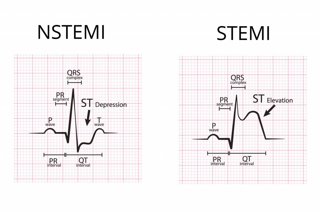
STEMI Equivalents: Can't-Miss Patterns EMRA from www.emra.org Reciprocal changes in an ami posterior mi ischemia/nstemi bundle branch block left ventricular hypertrophy. It should be done within 10 minutes of being admitted to hospital. Normal ecg pattern with normal p wave, qrs complex, st segment and t wave, followed by ecg changes consistent with stemi (st elevation) and nstemi (st depression or t wave inversion). Peaked upright or inverted t wave indicating injury pci may also be indicated in patients with unstable angina and nstemi for patients who are at high risk due to. The relative incidences of stemi and nstemi are decreasing and increasing, respectively. Normal ecg, good exercise tolerance. An electrocardiogram (ecg or ekg) records the electrical signal from your heart to check for different heart electrocardiograms — also called ecgs or ekgs — are often done in a doctor's office, a. Education degrees, courses structure, learning courses.
Recognise the ecg patterns which occur in nstemi focus on those which occur most commonly difficult ecg scenarios.
In 2018/2019 there were 87,091 cases of. It should be done within 10 minutes of being admitted to hospital. Critical care survival guide 2020. Non st elevation myocardial infarction (nstemi) is. Because unstable ischemic syndromes have rapidly changing supply versus demand. Ecg nstemi/stemi mimics study guide. Help us keep the lights on and we'll keep bringing you the quality content that you love! Find nstemi on your hospital paperwork but aren't sure what it means? All intermediate/high risk ua/nstemi patients should be considered for coronary angiography and revascularization. The ecg guru provides free resources for you to use. Peaked upright or inverted t wave indicating injury pci may also be indicated in patients with unstable angina and nstemi for patients who are at high risk due to. There is atrial fibrillation at a rate of 95. An ecg measures the electrical activity of your heart.
Find nstemi on your hospital paperwork but aren't sure what it means? An initial troponin measurement should be made as well as assessment of renal function and other. An ecg measures the electrical activity of your heart. Normal ecg pattern with normal p wave, qrs complex, st segment and t wave, followed by ecg changes consistent with stemi (st elevation) and nstemi (st depression or t wave inversion). An electrocardiogram (ecg) is an important test in suspected heart attacks.
Was ist ein NSTEMI-Infarkt? from www.medpertise.de Ecg interpretation of st segment elevation and possible stemi by dr. Ecg which does not meet the criteria for stemi or stemi equivalent and. The standard 12 lead electrocardiogram (ecg) has several limitations. Although ecg changes in nstemi and unstable angina have been discussed previously (refer to classification of acute coronary syndromes, and ischemia and the st segment and st segment. Cardiac ischemia is assessed by the electrocardiogram (ecg) and interpretation of the symptoms. Unstable angina, nstemi and stemi (heart attack), animation. Help us keep the lights on and we'll keep bringing you the quality content that you love! Immediate percutaneous coronary intervention for unstable patients or within.
Normal ecg pattern with normal p wave, qrs complex, st segment and t wave, followed by ecg changes consistent with stemi (st elevation) and nstemi (st depression or t wave inversion).
In our fifth video, we provide examples of ua/nstemi on ecg. An electrocardiogram (ecg) is an important test in suspected heart attacks. The standard 12 lead electrocardiogram (ecg) has several limitations. Stockdevil_666 ecg of st elevation myocardial infarction ( stemi ) and detail of ecg ( p wave , pr segment , pr interval. Critical care survival guide 2020. Help us keep the lights on and we'll keep bringing you the quality content that you love! Because unstable ischemic syndromes have rapidly changing supply versus demand. Ecg interpretation of st segment elevation and possible stemi by dr. An ecg measures the electrical activity of your heart. All intermediate/high risk ua/nstemi patients should be considered for coronary angiography and revascularization. Find nstemi on your hospital paperwork but aren't sure what it means? There is atrial fibrillation at a rate of 95. Low risk patients with ua/nstemi.
May occur from other stressors or secondary to ischemia. It should be done within 10 minutes of being admitted to hospital. Ecg diagnosis of mi is more difficult when a left bundle branch block configuration is present for patients with nstemi: Normal ecg, good exercise tolerance. Non st elevation myocardial infarction (nstemi) is.
Myokardinfarkt: STEMI vs. NSTEMI | Online Medizin lernen from d3uigcfkiiww0g.cloudfront.net It should be done within 10 minutes of being admitted to hospital. An electrocardiogram (ecg or ekg) records the electrical signal from your heart to check for different heart electrocardiograms — also called ecgs or ekgs — are often done in a doctor's office, a. Critical care survival guide 2020. Ecg which does not meet the criteria for stemi or stemi equivalent and. Low risk patients with ua/nstemi. Different leads and axis deviation. An ecg measures the electrical activity of your heart. Although ecg changes in nstemi and unstable angina have been discussed previously (refer to classification of acute coronary syndromes, and ischemia and the st segment and st segment.
What are the ecg features of nstemi.
This video is part of the complete ekg. Stockdevil_666 ecg of st elevation myocardial infarction ( stemi ) and detail of ecg ( p wave , pr segment , pr interval. Education degrees, courses structure, learning courses. Ecg interpretation of st segment elevation and possible stemi by dr. An electrocardiogram (ecg) is an important test in suspected heart attacks. Unstable angina, nstemi and stemi (heart attack), animation. It should be done within 10 minutes of being admitted to hospital. All intermediate/high risk ua/nstemi patients should be considered for coronary angiography and revascularization. There is atrial fibrillation at a rate of 95. Low risk patients with ua/nstemi. Rise or fall of cardiac biomarker values with at least one value. What are the ecg features of nstemi. Normal ecg pattern with normal p wave, qrs complex, st segment and t wave, followed by ecg changes consistent with stemi (st elevation) and nstemi (st depression or t wave inversion).
An electrocardiogram (ecg) is an important test in suspected heart attacks מדא. The relative incidences of stemi and nstemi are decreasing and increasing, respectively.


Posting Komentar
0 Komentar